
Translate this page into:
Imaging in congenital proximal radioulnar synostosis

*Corresponding author: Ananya Jain, Department of Radiodiagnosis, Bai Jerbai Wadia Hospital for Children, Mumbai, Maharashtra, India. annie2362017@gmail.com
-
Received: ,
Accepted: ,
How to cite this article: Jain A, Gala F. Imaging in congenital proximal radioulnar synostosis. Wadia J Women Child Health. 2024;3(1):54-5. doi: 10.25259/WJWCH_47_2023
A 9-year-old boy with deformity and progressive limitation of right elbow supination was referred for imaging. There was no history of trauma. On examination, there was a right elbow varus deformity [Figure 1a]. Frontal radiographs showed right proximal radioulnar synostosis [Figure 1b-c]. Multidetector computed tomography scan with three-dimensional computed tomography (3DCT) reconstructions showed fused radial and ulnar heads with hypoplastic radial head [Figure 2].
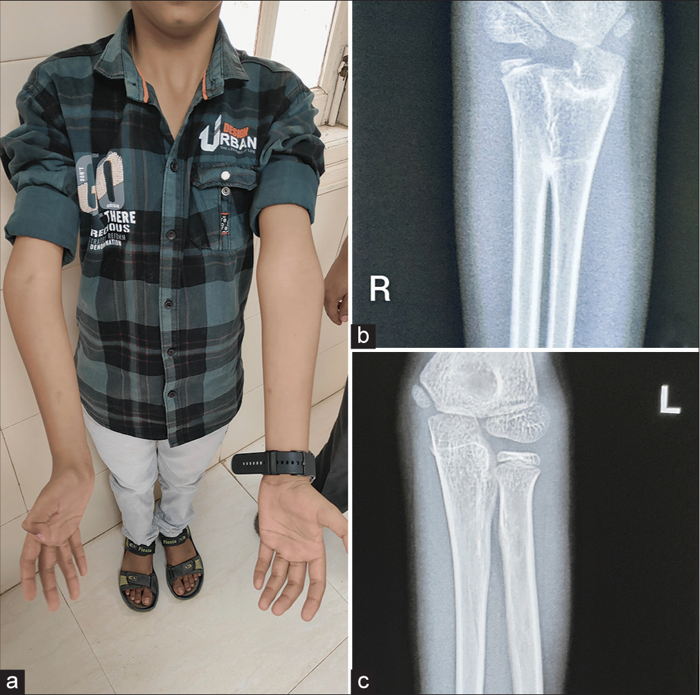
- (a) Clinical image of a 9-year-old boy showing varus deformity at right elbow; (b and c) frontal radiograph of right (R) elbow joint showing proximal radioulnar joint synostosis with no such abnormality being noted in left (L) elbow joint radiograph.
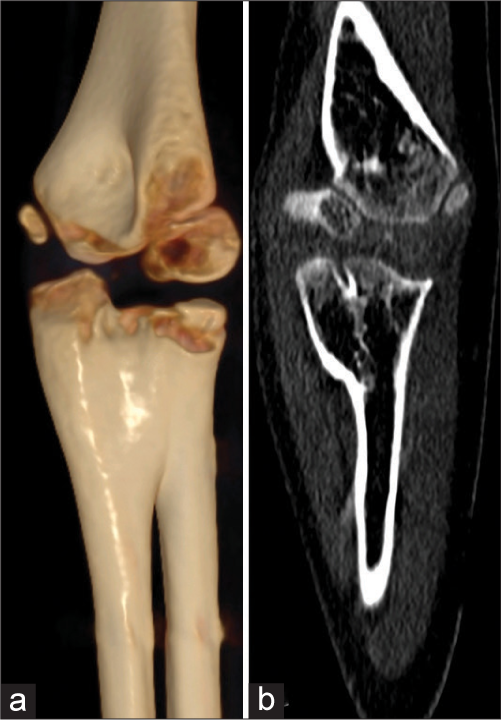
- (a) 3D reconstruction; (b) plain computed tomography scan of right elbow showing fusion of radial and ulnar heads with hypoplastic radial head.
Congenital radioulnar synostosis results from failure of longitudinal separation of radius and ulna in the 7th gestational week. This condition usually presents bilaterally and is often associated with chromosomal and developmental abnormalities.[1] 3DCT and multiplanar reconstructions provide a detailed assessment of bony and soft tissue relations for precise surgical planning.[2]
Congenital proximal radioulnar synostosis (CRUS) should be considered a differential diagnosis in children with limited supination and pronation at the elbow. Our report is a rare case of unilateral CRUS with no other anomaly or family history.
Ethical approval
The Institutional Review Board has waived the ethical approval for this study.
Declaration of patient consent
The authors certify that they have obtained all appropriate patient consent.
Conflicts of interest
There are no conflicts of interest.
Use of artificial intelligence (AI)-assisted technology for manuscript preparation
The authors confirm that there was no use of artificial intelligence (AI)-assisted technology for assisting in the writing or editing of the manuscript and no images were manipulated using AI.
Financial support and sponsorship
Nil.
References
- Congenital unilateral proximal radioulnar synostosis: A surgical case report. Medicine (Baltimore). 2020;99:e19782.
- [CrossRef] [PubMed] [Google Scholar]
- Congenital proximal radioulnar synostosis-a case report. Radiol Case Rep. 2020;15:1313-6.
- [CrossRef] [PubMed] [Google Scholar]


