
Translate this page into:
Sampling makes a difference in surgical pathology
*Corresponding author: Dr. Leena Patwardhan, Consultant Pathologist, Bai Jerbai Wadia Hospital for Children, Mumbai, Maharashtra, India. leenapatwardhan68@gmail.com
-
Received: ,
Accepted: ,
How to cite this article: Sharma S, Patwardhan L, Mamtora D. Sampling makes a difference in surgical pathology. Wadia J Women Child Health 2022;1(1):40-1.
We report an interesting case wherein we received a lymph node [Figure 1] for processing; the lymph node showed reactive lymphadenitis; however, at the edge; there was a small attached connective tissue. That connective tissue harboured granulomatous tuberculous inflammation, clinching the diagnosis of Tuberculosis, which otherwise would have been missed. Therefore, if complete good material obtained by surgical biopsies is sent to one laboratory for diagnostic review, it will aid in diagnosis and reduce chances of error.
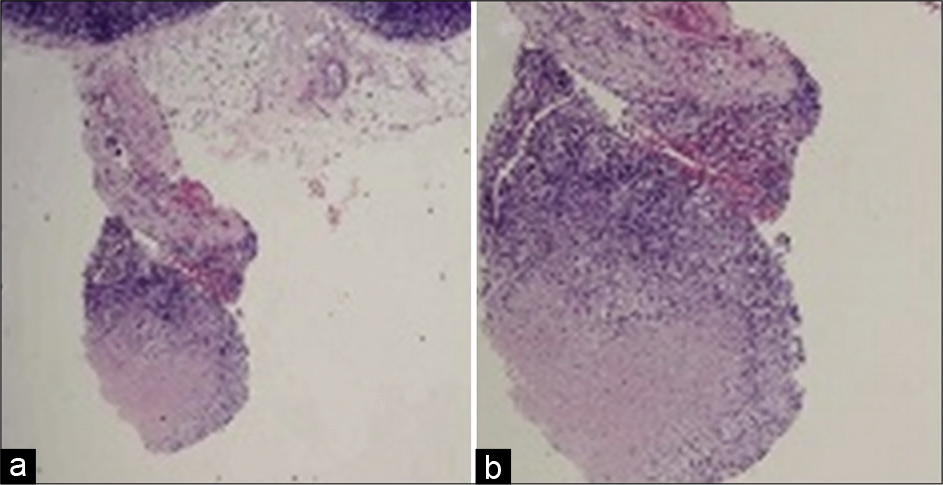
- (a) Reactive lymph node showing connective tissue at edge. (b) Connective tissue edge showing granulomatous lymphadenitis.
Single lymph-node [Figure 2] for histopathological examination. It is important to know value of correct sampling. Above image Figure 2a shows a small section taken for examination and Immuno-Histo-Chemistry (IHC); which shows a reactive process in the lymph node. Figure 2b shows a different section taken from the same lymphnode for examination and IHC; showing presence of Mantle zone lymphoma.
![(a) A small section of lymph node for examination and IHC (immunohistochemistry). (b) Section taken from the same lymphnode for examination and IHC; showing presence of Mantle zone lymphoma. (Picture taken from google for illustrating purpose). [1]](/content/147/2022/1/1/img/WJWCH-1-040-g002.png)
- (a) A small section of lymph node for examination and IHC (immunohistochemistry). (b) Section taken from the same lymphnode for examination and IHC; showing presence of Mantle zone lymphoma. (Picture taken from google for illustrating purpose). [1]
Declaration of patient consent
Patient’s consent not required as patients identity is not disclosed or compromised.
Financial support and sponsorship
Nil.
Conflicts of interest
There are no conflicts of interest.
References
- Accurate diagnosis of lymphoma on whole-slide histopathology images using deep learning. NPJ Digit Med. 2020;3:63.
- [CrossRef] [PubMed] [Google Scholar]


