
Translate this page into:
Refractory or recurrent iron deficiency anemia: Explore before you prick

*Corresponding author: Ritika Khurana, Department of Pediatric Hematology and Oncology, Bai Jerbai Wadia Hospital for Children, Mumbai, India. ritangel247@gmail.com
-
Received: ,
Accepted: ,
How to cite this article: Khurana R, Kanvinde P, Mudaliar S. Refractory or recurrent iron deficiency anemia: Explore before you prick. Wadia J Women Child Health. 2024;3(1):52-3. doi: 10.25259/WJWCH_49_2023
A 10-year-old well grown boy, first by birth order, born of non-consanguineous marriage was referred for recurrent microcytic, hypochromic anemia temporarily responding to iron supplementation. He had multiple bluish patches on the sole of foot, calf muscle, and abdomen [Figure 1] and bony enlargement of scapula [Figure 2]. Weight and height was at 10th centile. Stool for occult blood was positive. Endoscopy revealed similar lesions in GI mucosa. His anemia improved and lesions reduced in size on iron supplementation and propranolol.
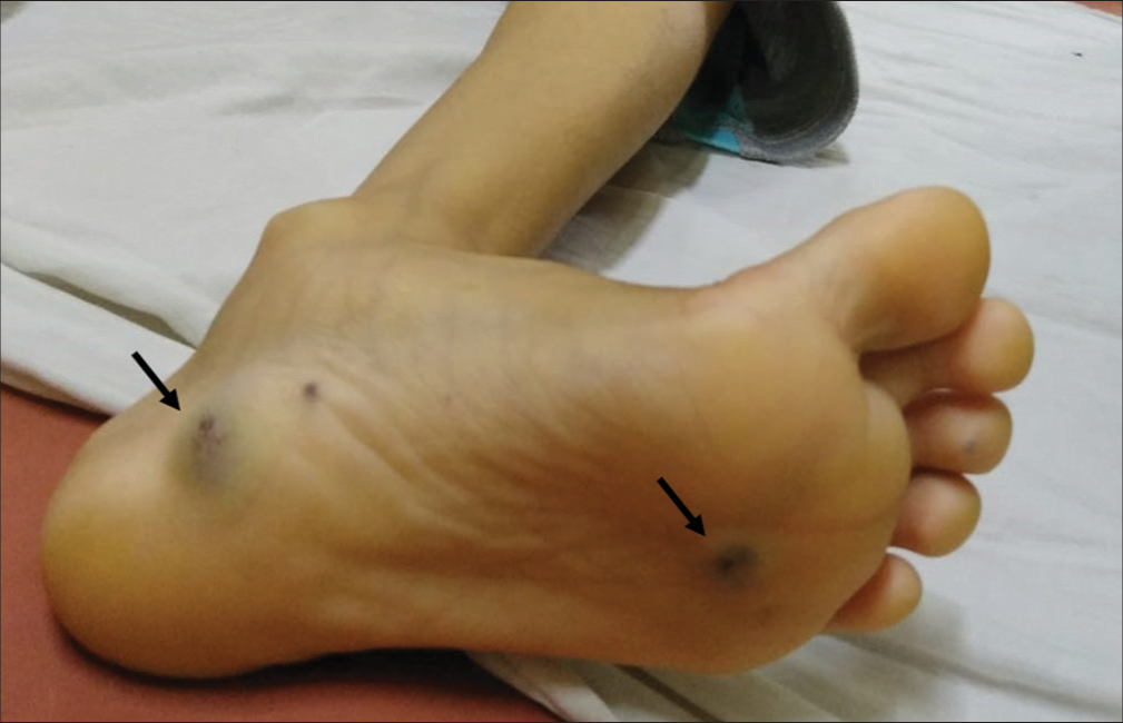
- Nevus present as soft, non-mobile, dark blue, compressible papules, as shown by black arrows.
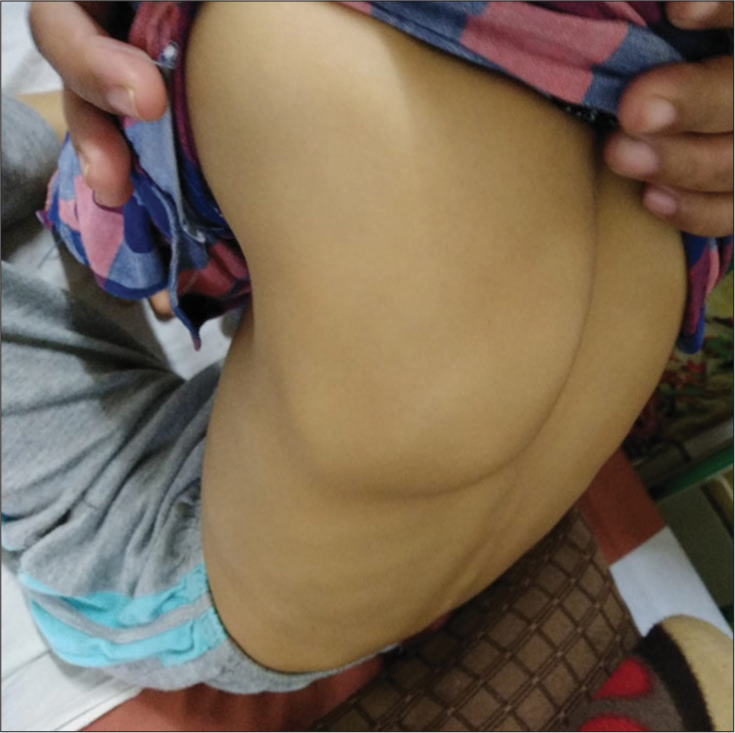
- Bony deformity.
Blue rubber bleb nevus syndrome is a rare disorder with vascular malformations in skin, soft tissue, and gastrointestinal tract inherited in autosomal dominant fashion. Most patients present with chronic refractory microcytic, hypochromic anemia, and rarely massive GI bleed can occur. Bony abnormalities occur secondary to pressure from adjacent hemangiomas or due to hypervascularity. Gastrointestinal lesions may require antiangiogenic agents or endoscopic management. For all refractory anemias, extensive examination and search for site of blood loss should be done. In disorders such as blue rubber bleb nevus syndrome, targeted treatment strategies can be planned depending on the extent of involvement.
Ethical approval
Institutional Review Board approval is not required.
Declaration of patient consent
The authors certify that they have obtained all appropriate patient consent.
Conflicts of interest
There are no conflicts of interest.
Use of artificial intelligence (AI)-assisted technology for manuscript preparation
The authors confirm that there was no use of artificial intelligence (AI)-assisted technology for assisting in the writing or editing of the manuscript and no images were manipulated using AI.
Financial support and sponsorship
Nil.


