
Translate this page into:
PHACE syndrome
-
Received: ,
Accepted: ,
How to cite this article: Rakshpaul S, Chauhan N. PHACE syndrome. Wadia J Women Child Health 2022;1(1):38-9.
Twenty two-days-old female, second child of a non-consanguineous marriage born at 38 week, 3500 g weight, appropriate for Gestational age with no significant Antenatal/intrapartum/ postnatal period, was brought to High-Risk OPD with complaints of gradually increasing rash with excoriations on the left side of the face, including upper lip, nostril, temple, and neck [Figure 1a] noticed after a few days of birth.
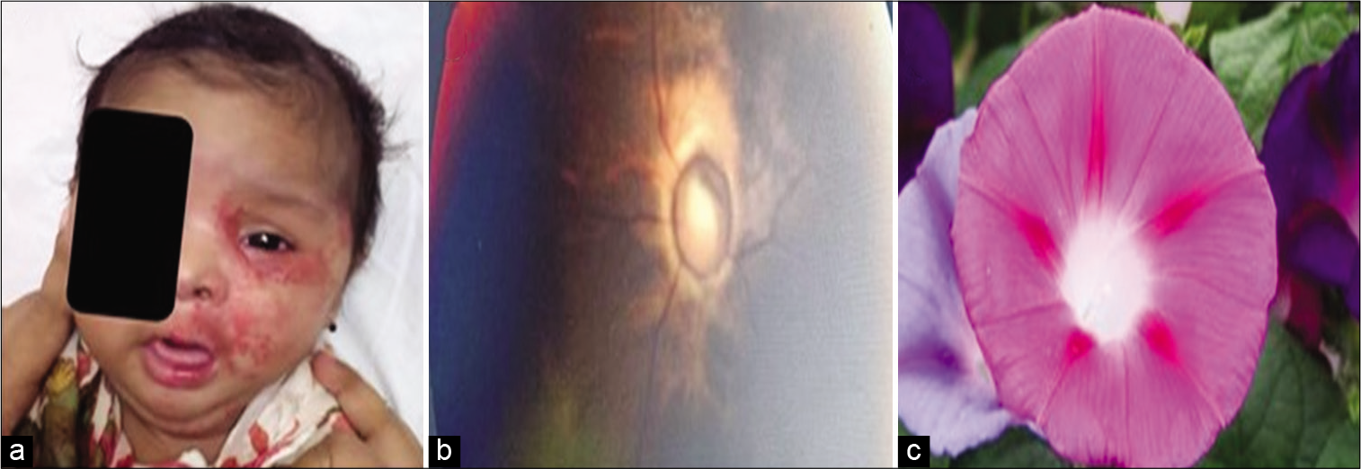
- (a) Haemangioma, (b) fundus showing enlarged disc with vasculature, (c) like morning glory.
For a complete PHACE survey, -(P)osterior fossa abnormality; (H)emangioma of the face, neck and scalp; anatomical anomalies of cerebral or cervical (A)rteries; (C)ardiac anomalies/ (C)oarctation of aorta; (E)ye abnormalities. If (S)ternal anomalies are present then it is termed as “PHACES” syndrome.[1] MRA brain was done which revealed left midbrain hypoplasia, prominent and tortuous basilar artery, small caliber fetal origin of a right posterior cerebral artery, prominent left supraclinoid internal carotid artery, and proximal, middle cerebral artery. Fundus examination was done which is as shown in [Figure 1b]. The lesion has got its name from the flower morning glory in [Figure 1c]. Echocardiogram done was normal.
Presently, baby is on regular follow-up on propranolol showing normal growth parameters and normal INFANIB at 9 months.
Acknowledgments
Neonatology Team, B. J. Wadia Hospital.
Declaration of patient consent
The authors certify that they have obtained all appropriate patient consent.
Financial support and sponsorship
Nil.
Conflicts of interest
There are no conflicts of interest.
References
- Consensus statement on diagnostic criteria for PHACE syndrome. Pediatrics. 2009;124:1447-56.
- [CrossRef] [PubMed] [Google Scholar]


